Millions of people worldwide suffer from neurodegenerative diseases such as Alzheimer’s disease (AD), Parkinson’s disease (PD), and amyotrophic lateral sclerosis (ALS) due to the gradual loss of neurons. These diseases often share a major contributing factor: mitochondrial dysfunction, which leads to cellular energy imbalance.
Mitochondria are responsible for generating most of the cell’s ATP, and when they do not function properly, neurons experience metabolic stress, reduced activity, and eventual degeneration.
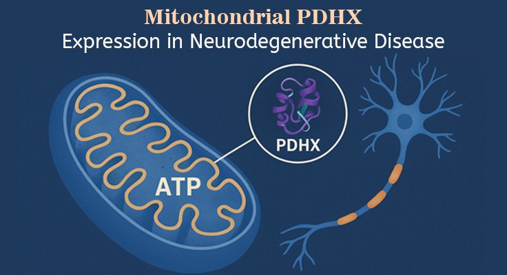
The pyruvate dehydrogenase complex (PDC) is a key mitochondrial enzyme system that converts pyruvate into acetyl-CoA (a crucial fuel for the cell). The protein PDHX helps hold the structure together within the pyruvate dehydrogenase complex (PDC) and ensures the complex functions efficiently. PDHX imbalance can weaken the entire PDC and reduce energy production in neurons.
Researchers rely on tools like rabbit anti human PDHX antibodies to measure and visualize PDHX. The goal is to understand the contribution of metabolic failures to disease progression.
PDHX Function and Neuronal Energy Metabolism
The pyruvate dehydrogenase complex (PDC) links glycolysis to the tricarboxylic acid (TCA) cycle and enables efficient production of ATP. Acting as a structural scaffold, PDHX anchors the E3 subunit to the E2 core, which ensures:
- Proper assembly of the PDC
- Efficient enzymatic activity
- Stable conversion of pyruvate into acetyl-CoA
Neurons heavily rely on oxidative phosphorylation, and even small disruptions in PDC function can lead to energy deficits. Reduced PDHX levels destabilize the PDC, which lowers ATP production and causes metabolic stress.
This leads to the following metabolic consequences:
- Pyruvate and lactate accumulation
- Impaired NADH/NAD⁺ balance
- Increased neuronal susceptibility to stress and injury
PDHX integrity is essential for neuronal survival, synaptic function, and overall mitochondrial efficiency.
Researchers use rabbit anti human PDHX antibodies to localize and quantify PDHX. This provides insights into mitochondrial function and metabolic failure.
PDHX Antibodies Applications in Neurodegeneration Research
| Technique | Purpose | Recommended Antibody Concentration |
| Western Blot | Quantitative assessment of PDHX in tissue lysates | 0.5–2 µg/mL |
| Immunocytochemistry | Visualization of PDHX subcellular localization in cultured neurons | 5–20 µg/mL |
| Immunohistochemistry | Mapping PDHX in frozen or paraffin-embedded human brain tissue | 5–20 µg/mL |
| ELISA | High-throughput quantitative PDHX detection | 0.05–2 µg/mL |
Experimental Considerations for Researchers
Selection of Antibody and Validation
Make sure that the rabbit anti human PDHX antibodies have been validated for the intended application.
Antibody specificity must be confirmed using positive and negative controls. Also consider batch-to-batch variability and cross-reactivity with related proteins, as both can influence reproducibility.
Sample Preparation
When it comes to preparing samples for Western Blot and ELISA, use high-quality tissue or cell lysates with protease inhibitors. This helps in preserving the integrity of the protein, which is essential for accurate detection.
Immunocytochemistry and Immunohistochemistry require proper fixation and permeabilization conditions. This helps retain subcellular localization and access to the antibody.
Use fresh or properly stored frozen tissue to ensure accurate detection of PDHX.
Quantification and Imaging
When it comes to immunocytochemistry or immunohistochemistry, measure fluorescence intensity or signal area to quantify PDHX.
Getting a Complete Picture
PDHX levels are often influenced by the following factors:
- Cell type
- Metabolic state
- Disease state
Combining PDHX measurements with mitochondrial activity assays provides a complete picture.
Mechanistic Insights
| Key Mechanistic Pathway | Description/Impact |
| Energy Production Deficits | Reduces PDC activityLowers acetyl-CoA, NADH, FADH2Impairs ATP production and neuronal signaling |
| Metabolic Imbalance & Oxidative Stress | Causes pyruvate/lactate buildupIncreases reactive oxygen species (ROS)Leads to energy deficits and oxidative stress |
| Regulation of Mitochondrial Dynamics | Affects mitochondrial morphologyDisrupts fission/fusion balanceImpacts axonal transport and synaptic function |
| Interaction with Neurodegenerative Pathways | Worsens protein aggregationImpairs autophagyLinks metabolic dysfunction to disease progression |
Conclusion
PDHX plays a critical role in maintaining PDC stability and neuronal energy homeostasis. PDHX disruptions decrease production, alter mitochondrial dynamics, and increase oxidative stress, leading to neuronal degeneration.
Mitochondrial dysfunction is a central contributor to neurodegenerative diseases. Understanding PDHX regulation and its impact on mitochondrial function may help develop therapeutic strategies to restore neuronal metabolism and mitigate neurodegeneration.
Also Read: Prion Strain Variability and Its Impact on Disease Phenotype and Progression